
Imaging Systems: Gel Documentation, Fluorescence Imaging and Live-Cell Imaging
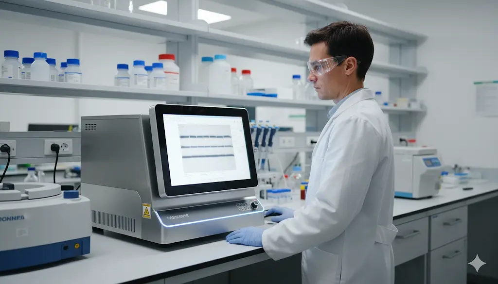
Laboratory imaging systems play a central role in molecular biology, cell biology, proteomics, genomics, pharmaceutical research, and biomedical laboratories. These instruments are used to capture, document, and analyze visual data from gels, membranes, fluorescent samples, and live cells, helping researchers move from simple image capture to quantitative biological analysis. Depending on the workflow, a lab may use a gel documentation system, fluorescence imaging system, chemiluminescence imager, or live-cell imaging platform.
For organizations comparing laboratory imaging systems, gel documentation equipment, fluorescence imagers, live-cell imaging systems, and used imaging equipment, the best platform depends on sample type, detection method, sensitivity, resolution, imaging speed, software, automation, and compatibility with existing assays. A basic gel documentation system may be ideal for routine DNA or protein gels, while a live-cell platform may need environmental control, time-lapse capability, and automated image analysis. Choosing the right imaging system can improve data quality, reduce manual interpretation, and make research workflows more efficient.
What Is a Laboratory Imaging System?
A laboratory imaging system is an instrument designed to capture and analyze visual information from biological or chemical samples. These systems may use visible light, ultraviolet illumination, fluorescence, chemiluminescence, or transmitted light depending on the application. The resulting images can be used for documentation, comparison, quantification, and experimental analysis.
Many modern imaging systems combine sensitive cameras, controlled illumination, optical filters, and analysis software in one platform. This allows laboratories to standardize how images are captured and reduce variation between operators. Higher-end systems may also support automated focusing, multiple fluorescent channels, time-lapse imaging, and quantitative measurements.
The term covers a broad range of instruments, so it is important to distinguish between systems designed for gels and blots, fluorescence-based assays, and live-cell applications. Each category has different performance requirements and may use very different optics, cameras, and environmental controls.
Gel Documentation Systems
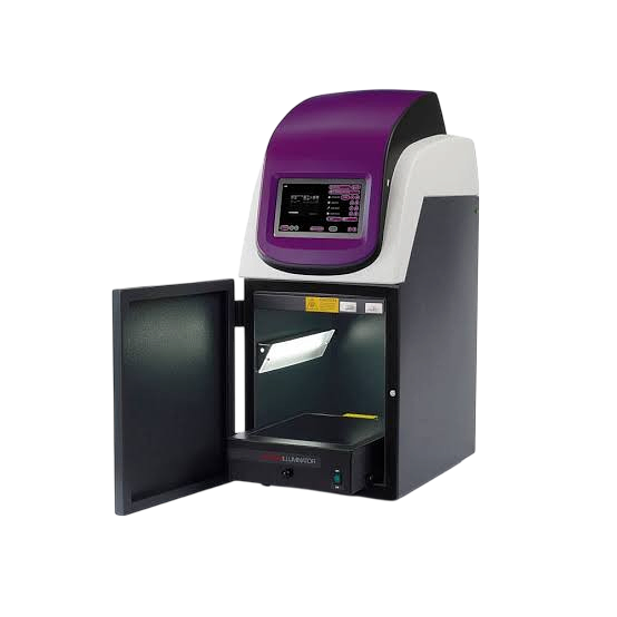
Gel documentation systems are used to capture images of electrophoresis gels after DNA, RNA, or protein separation. Depending on the stain and application, these systems may use UV, blue-light, or visible-light illumination to visualize bands and generate a permanent digital record.
They are commonly used in PCR workflows, cloning, molecular biology, genomics, and protein research. Digital gel documentation systems can replace manual photography and make it easier to compare band intensity, estimate molecular size, document experimental results, and store images for future reference.
When evaluating a gel imager, camera resolution, illumination type, filter compatibility, field of view, and software should all be considered. Laboratories working with sensitive nucleic acids may also prefer blue-light systems when minimizing UV exposure is important.
Fluorescence Imaging Systems
Fluorescence imaging systems are designed to detect fluorescent labels and dyes across one or more wavelength channels. These systems are used for Western blots, gels, membranes, microplates, tissue samples, and other fluorescent assays where signal intensity and channel separation are important.
Compared with basic gel documentation equipment, fluorescence imagers often provide greater sensitivity, wider dynamic range, and more advanced optical filtering. They may also support multiplex experiments, allowing multiple targets to be detected and quantified from the same sample.
These systems are widely used in protein research, molecular biology, cell biology, biomarker studies, and pharmaceutical development. Software for background correction, normalization, band quantification, and image comparison can significantly increase the analytical value of the instrument.
Chemiluminescence and Western Blot Imaging
Chemiluminescence imaging systems are commonly used for Western blot detection and other assays that generate light through a chemical reaction. Rather than relying on external illumination, the system uses a highly sensitive camera to detect low-level light emitted by the sample.
These platforms are especially important in protein analysis because weak bands may require longer exposures and high detector sensitivity. Digital chemiluminescence imaging can also simplify workflows compared with film-based methods by allowing users to adjust exposure, archive images, and perform quantitative analysis electronically.
Many modern systems combine chemiluminescence with fluorescence and visible-light imaging in one unit. This can make a multi-mode imager a strong choice for labs that handle several protein and molecular biology workflows.
Live-Cell Imaging Systems
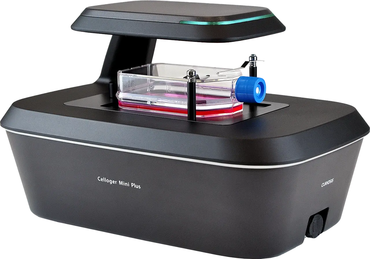
Live-cell imaging systems are designed to observe living cells over time without removing them from controlled culture conditions. These platforms may be integrated with incubators or include their own temperature, humidity, and gas control to maintain an environment suitable for long-term imaging.
Live-cell systems are widely used for cell proliferation studies, wound-healing assays, apoptosis research, cell migration, drug screening, stem cell research, and oncology applications. Time-lapse imaging allows researchers to monitor biological changes continuously instead of relying only on endpoint measurements.
More advanced systems can automate image capture across multiple wells or positions, track cells over time, and quantify changes in confluence, morphology, fluorescence, or movement. This level of automation can significantly improve throughput in cell-based research and pharmaceutical screening.
Gel Documentation vs. Fluorescence vs. Live-Cell Imaging
A gel documentation system is generally designed for static samples such as DNA gels, protein gels, or stained membranes. A fluorescence imaging system expands the workflow by detecting fluorescent signals with greater sensitivity and supporting more complex assays.
A live-cell imaging system is fundamentally different because the sample remains alive throughout the experiment. These systems must therefore combine imaging with environmental control and repeated acquisition over time.
Laboratories should select equipment based on the sample and experiment rather than simply choosing the system with the greatest number of features. A live-cell platform may be unnecessary for routine gel imaging, while a basic gel doc system will not support long-term cellular imaging or automated cell analysis.
Common Applications for Laboratory Imaging Systems
Laboratory imaging equipment is used across a wide range of life science and analytical workflows.
Common applications include:
- DNA and RNA gel documentation
- Protein gel imaging
- Western blot detection
- Fluorescence imaging
- Chemiluminescence detection
- Cell proliferation studies
- Live-cell time-lapse imaging
- Drug screening
- Cell migration and wound-healing assays
- Biomarker and protein analysis
These systems are commonly found in academic research, biotechnology, pharmaceutical development, clinical research, and contract laboratories. Imaging quality directly affects how confidently researchers can interpret experimental results, so sensitivity, resolution, and software performance are important purchasing considerations.
What to Consider When Purchasing an Imaging System
When purchasing a used laboratory imaging system, the first step is determining exactly what sample types and detection methods will be used. A system optimized for fluorescent Western blots may not be appropriate for live-cell imaging, and a live-cell instrument may be unnecessarily complex for routine gel documentation.
Important considerations include:
- Imaging mode
- Camera sensitivity
- Resolution
- Fluorescence channels
- Chemiluminescence capability
- UV or blue-light illumination
- Live-cell environmental control
- Automated focusing
- Time-lapse capability
- Analysis software
- Computer and software compatibility
- Overall instrument condition
Camera and illumination condition are especially important. Aging light sources, damaged filters, weak detectors, or missing optical components can significantly reduce performance even if the instrument powers on normally.
Software should also be reviewed carefully, particularly with advanced imaging platforms. A complete system with the original computer, software, license, camera, filters, and accessories can offer much better value than a lower-priced unit missing key components.
Recommended Laboratory Imaging Systems
Below are several widely recognized gel documentation, fluorescence imaging, and live-cell imaging systems used in molecular biology, protein research, and cell-based laboratories.
Bio-Rad ChemiDoc MP Imaging System – A widely used multi-mode imaging platform for chemiluminescence, fluorescence, and gel documentation. It is commonly used in Western blotting, protein analysis, and molecular biology labs that need sensitive digital imaging and quantitative software.
Azure Biosystems cSeries Imaging Systems – Flexible imaging platforms used for chemiluminescent Western blots, fluorescence imaging, and gel documentation. These systems are popular in research labs that need multiple detection modes from a single workstation.
LI-COR Odyssey Imaging Systems – Well-known fluorescence imaging systems used for quantitative Western blotting and multiplex protein detection. They are especially valuable in labs that rely heavily on near-infrared fluorescence workflows.
Thermo Scientific iBright Imaging Systems – Integrated imaging platforms designed for Western blotting and gel documentation. These systems combine automated image capture with software tools for band detection, normalization, and analysis.
Sartorius Incucyte Live-Cell Analysis Systems – Widely used live-cell imaging platforms designed for time-lapse imaging and automated cell analysis inside a controlled incubation environment. Incucyte systems are commonly used in cell biology, oncology, drug discovery, and proliferation studies.
Agilent BioTek Cytation Cell Imaging Multi-Mode Readers – Multi-function systems that combine imaging with microplate-based detection. These platforms are useful for laboratories running cell-based assays, fluorescence imaging, and higher-throughput screening workflows.
Why Used Imaging Systems Can Offer Strong Value
New gel documentation systems, fluorescence imagers, and live-cell imaging platforms can be expensive, especially when advanced cameras, optical filters, incubation control, and software are included. A tested used system can provide access to proven imaging technology at a lower acquisition cost, making it attractive for biotech startups, academic laboratories, CROs, and facilities expanding existing imaging capacity.
Used systems can be especially valuable when a laboratory already uses the same manufacturer or platform family. Existing methods, software workflows, accessories, imaging protocols, and operator experience may remain compatible, reducing setup time and retraining.
Condition and completeness should be evaluated carefully. A well-maintained imaging system with a functioning camera, intact optical path, compatible software, and complete accessories may provide better value than a newer but incomplete platform. For live-cell systems, environmental controls, stage movement, autofocus, and incubation performance should also be checked.
Choosing the Right Imaging System
The right laboratory imaging system depends on the sample type, signal, throughput, and level of analysis required. A molecular biology lab focused on DNA gels may need only a reliable gel documentation system, while a protein lab may benefit from fluorescence and chemiluminescence capability. A cell biology group performing long-term experiments may need a live-cell imaging platform with automated acquisition and environmental control.
It is also important to consider how the instrument fits into the existing workflow. Software compatibility, plate or sample format, imaging speed, and analysis tools can have as much impact on usability as the camera itself. By comparing detection modes, optical performance, automation, software, and overall condition, laboratories can select used imaging equipment that supports current research needs without paying for unnecessary features.
Looking for Laboratory Imaging Systems?
Explore our inventory of used gel documentation systems, fluorescence imagers, chemiluminescence systems, live-cell imaging platforms, and molecular biology equipment, or consign your surplus instruments to connect with qualified buyers.
info@rebio.com