
Western blot systems are widely used in molecular biology, biotechnology, pharmaceutical research, proteomics, and academic laboratories to identify and characterize specific proteins within complex biological samples. A complete Western blot workflow may involve protein electrophoresis, membrane transfer, antibody-based detection, chemiluminescent or fluorescent imaging, and quantitative analysis, making equipment selection important for both analytical performance and laboratory throughput. Modern platforms range from conventional transfer and imaging systems to highly automated instruments that combine multiple Western blotting steps into a single workflow.
Laboratories comparing Western blot equipment, protein transfer systems, Western blot imagers, chemiluminescence imaging systems, automated Western blot analyzers, and used protein analysis equipment should consider sample throughput, detection method, sensitivity, automation, software, consumables, and compatibility with existing protocols. A research group processing a few membranes each week may need very different equipment from a pharmaceutical or biotech lab running large numbers of protein assays. Choosing the right Western blot platform can reduce hands-on time, improve reproducibility, and make quantitative protein analysis significantly more efficient.
What Is a Western Blot System? 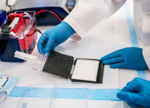
A Western blot system is used to detect specific proteins after they have been separated according to molecular size or other characteristics. In a traditional workflow, proteins are first separated by gel electrophoresis and then transferred onto a membrane, where antibodies are used to selectively identify the protein of interest. The resulting signal is detected using chemiluminescence, fluorescence, or another imaging method, producing data that can be used to compare protein expression between samples.
The term Western blot system can therefore describe several different types of equipment rather than one instrument. Protein transfer systems move proteins from gels to membranes, Western blot imaging systems capture the resulting signal, and automated platforms can integrate separation, immunodetection, and analysis into a single instrument. Understanding where a piece of equipment fits within this workflow is important when comparing systems because a transfer unit, imager, and automated analyzer perform different functions even though all belong to the Western blotting category.
Protein Transfer Systems
Western blot protein transfer systems move separated proteins from a polyacrylamide gel onto a membrane such as PVDF or nitrocellulose. This step is critical because the membrane becomes the surface on which antibody binding and final protein detection occur. Transfer quality can influence sensitivity, band sharpness, and overall reproducibility, particularly when working with proteins at very high or very low molecular weights.
Traditional wet transfer systems remain common, while semi-dry and rapid transfer platforms can reduce processing time and reagent consumption. Some laboratories prioritize flexibility and lower consumable costs, while higher-throughput facilities may prefer systems capable of completing multiple transfers quickly with standardized settings. When selecting a transfer unit, membrane size, gel format, transfer speed, cooling requirements, and compatibility with existing electrophoresis equipment should all be considered.
Western Blot Imaging and Detection Systems
After antibody labeling, a Western blot imaging system captures the signal produced by the membrane. Chemiluminescence remains one of the most common detection methods, while fluorescence imaging allows multiple proteins or targets to be detected using different fluorescent channels. Some imaging platforms also support visible-light, colorimetric, or gel-imaging applications, allowing one instrument to serve several molecular biology workflows.
Sensitivity and dynamic range are important when comparing imagers because weak protein signals can be difficult to quantify accurately on lower-performance systems. Modern digital Western blot imagers can also replace traditional film workflows, reducing manual development steps and making images easier to store, analyze, and compare. Software for band quantification, normalization, molecular-weight estimation, and image documentation can add substantial value, particularly in research environments where quantitative results are required.
Automated Western Blot Systems
Automated Western blot analyzers reduce or eliminate many of the manual steps associated with conventional gel-based Western blotting. Depending on the platform, the system may automate protein separation, immunodetection, washing, signal generation, and data analysis. This can reduce hands-on time and improve consistency between users while allowing laboratories to process more samples within the same working day.
Automation is particularly attractive in biotechnology, pharmaceutical development, biomarker research, and other environments where reproducibility and throughput are priorities. However, automated systems may use proprietary cartridges, capillaries, antibodies, or reagent kits, making consumable cost an important part of the purchasing decision. Laboratories should therefore compare total workflow cost and assay compatibility rather than evaluating instrument price alone.
Common Applications for Western Blot Systems
Western blot equipment is used across a wide range of protein research and biopharmaceutical workflows.
Common applications include:
- Protein expression analysis
- Antibody validation
- Biomarker research
- Cell signaling studies
- Protein phosphorylation analysis
- Drug discovery and development
- Biopharmaceutical research
- Protein characterization
- Disease research
- Academic and translational research
Western blotting is particularly useful when researchers need to confirm the presence of a specific protein while also obtaining information about its apparent molecular weight. It is frequently used alongside ELISA, flow cytometry, mass spectrometry, PCR, and other analytical techniques to provide complementary biological data. Because Western blotting can be applied to such a broad range of research areas, both conventional and automated systems remain widely used across life science laboratories.
Chemiluminescence vs. Fluorescence Detection 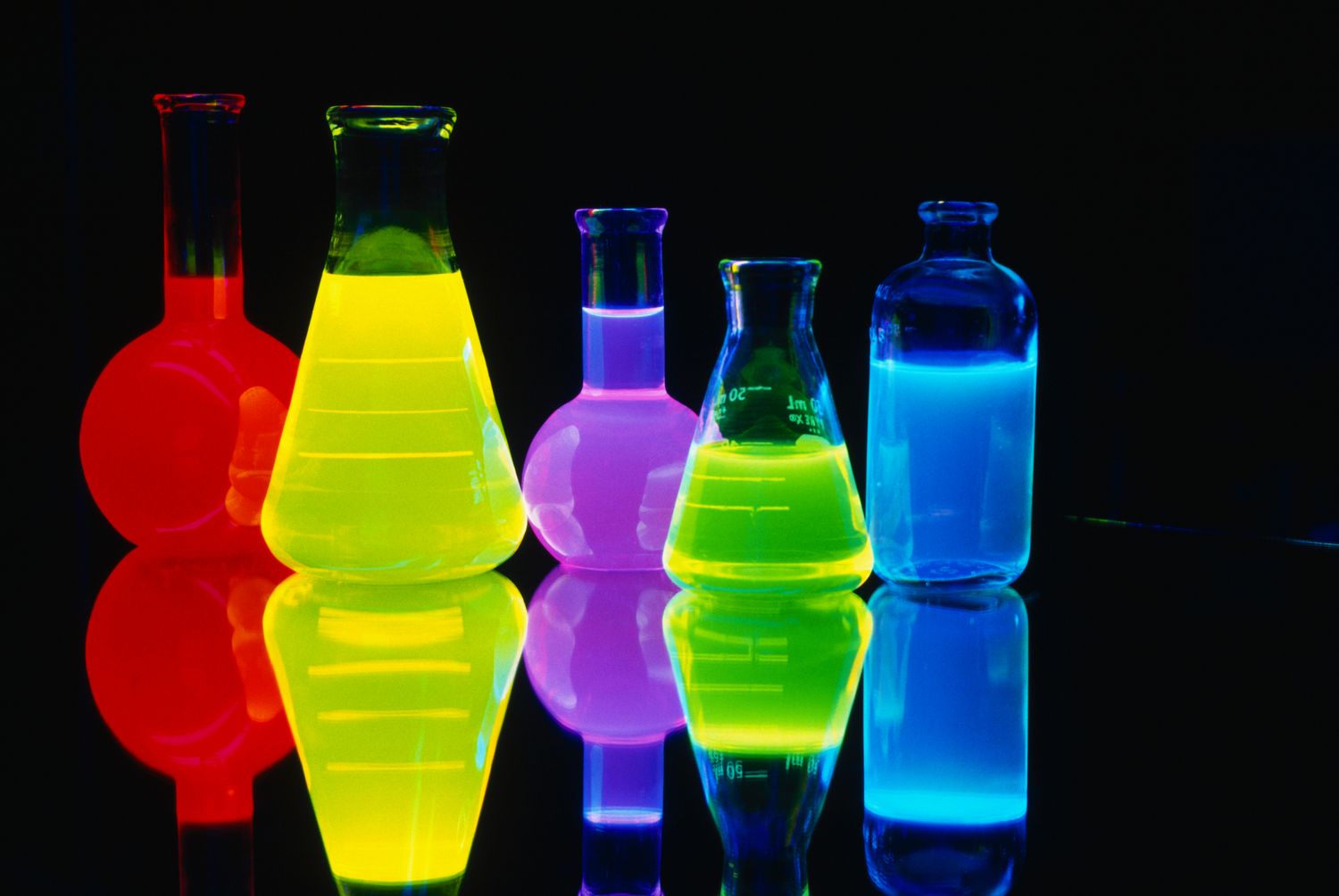
Chemiluminescent Western blot detection uses an enzyme-linked antibody reaction to generate light, which is then captured by an imaging system. It is widely used because of its sensitivity, established protocols, and compatibility with many commonly available antibodies and reagents. Laboratories already using traditional Western blot methods may find chemiluminescence the easiest workflow to maintain when upgrading from film to digital imaging.
Fluorescent Western blot detection uses fluorophore-labeled antibodies and can enable multiplex analysis, allowing multiple proteins to be detected on the same membrane when compatible dyes and imaging channels are used. Fluorescence can also offer a broad quantitative range and simplify normalization workflows. The best detection method depends on assay requirements, available antibodies, expected protein abundance, and whether multiplexing is important to the research program.
What to Consider When Purchasing a Western Blot System
When purchasing used Western blot equipment, the first step is determining whether the laboratory needs a transfer system, imaging platform, automated analyzer, or a combination of these components. A high-end imaging system will not replace a protein transfer unit in a conventional workflow, while an automated analyzer may eliminate several traditional steps entirely. Matching equipment to the existing laboratory protocol can prevent unnecessary purchases and workflow changes.
Important considerations include:
- Conventional or automated Western blot workflow
- Sample throughput
- Transfer method and gel compatibility
- Chemiluminescence or fluorescence detection
- Imaging sensitivity and dynamic range
- Multiplex detection capability
- Software and analysis tools
- Consumable and reagent availability
- Included accessories and computer hardware
- Overall system condition
Consumables deserve particular attention with automated Western blot platforms. Proprietary cartridges, capillaries, reagents, or detection kits may represent a significant portion of long-term operating costs, so continued availability should be confirmed before purchasing an older system. With imaging equipment, camera condition, illumination sources, filters, software licensing, and computer compatibility can be equally important.
Recommended Western Blot Systems
Below are several widely recognized Western blot analyzers, protein transfer systems, and imaging platforms used in biotechnology, pharmaceutical, molecular biology, and protein research laboratories. 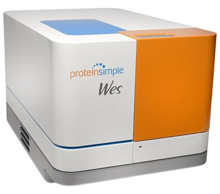
ProteinSimple WES Western Blot Protein Analyzer System – A highly automated Western blot protein analysis platform that uses capillary-based technology to streamline protein separation, immunodetection, and quantitative analysis. WES is especially useful for laboratories seeking to reduce the hands-on steps associated with traditional Western blotting while improving throughput and assay reproducibility.
Bio-Rad ChemiDoc MP Imaging System – A widely used digital Western blot imaging platform supporting chemiluminescent, fluorescent, and colorimetric detection. It is commonly found in molecular biology and protein research laboratories requiring sensitive imaging and quantitative analysis.
Bio-Rad Trans-Blot Turbo Transfer System – A popular rapid protein transfer system designed to move proteins from gels to membranes in significantly less time than many traditional wet transfer workflows. It is widely used for routine Western blotting in research and biotechnology laboratories.
Thermo Scientific iBlot 2 Gel Transfer Device – A compact dry-transfer platform designed for rapid protein transfer without the large buffer volumes required by conventional wet transfer systems. It is commonly used in laboratories seeking a simple and repeatable transfer workflow.
Azure Biosystems cSeries Imaging Systems – Digital imaging platforms used for chemiluminescent and fluorescent Western blot detection, gel imaging, and quantitative protein analysis. These systems are popular in research labs that need flexible imaging capability from a single workstation.
LI-COR Odyssey Imaging Systems – Well-known fluorescence-based Western blot imaging platforms frequently used for quantitative protein analysis and multiplex detection. They are especially popular in laboratories working with fluorescent antibody workflows and applications requiring broad dynamic range.
Why Used Western Blot Equipment Can Offer Strong Value
New Western blot analyzers and imaging systems can represent a significant laboratory investment, especially when automation, advanced cameras, fluorescence channels, and analysis software are included. Purchasing tested used equipment can provide access to established platforms at substantially lower acquisition cost, making advanced protein analysis more accessible to biotech startups, academic laboratories, CROs, and facilities expanding existing capacity. Used transfer systems and imagers can also provide an economical way to add backup capacity without changing the laboratory's established workflow.
Condition, completeness, and continued consumable support are more important than age alone. A complete Western blot system with the appropriate software, computer, trays, transfer accessories, cables, and documentation can be significantly easier to install and validate than an incomplete unit. Staying with an equipment family already used in the laboratory can also reduce training requirements and allow existing protocols, antibodies, imaging settings, and accessories to remain in use.
Choosing the Right Western Blot System
The best Western blot system depends on whether the priority is protein transfer, imaging, automation, throughput, sensitivity, or quantitative analysis. Conventional gel-and-membrane workflows provide substantial flexibility and allow laboratories to combine equipment from several manufacturers, while automated platforms such as the ProteinSimple WES can consolidate multiple steps and reduce technician involvement. Neither approach is ideal for every laboratory, so workflow requirements should guide equipment selection rather than simply choosing the most automated system available.
Laboratories performing frequent quantitative Western blots may prioritize sensitive digital imaging, broad dynamic range, and robust analysis software, while higher-throughput research groups may gain more value from automation. By comparing sample capacity, detection technology, consumables, software, workflow compatibility, and overall equipment condition, laboratories can select Western blot equipment that improves protein analysis while controlling acquisition and operating costs.
Looking for Western Blot & Protein Analysis Equipment?
Explore our inventory of used Western blot systems, protein analyzers, transfer systems, imaging equipment, and molecular biology instruments, or consign your surplus laboratory equipment to connect with qualified buyers.
info@rebio.com